The Sensitivity of Immunohistochemical Expression of p53 as an Indicator of the Malignant Potential of Gastric Hyperplastic Polyps: A Retrospective Study
DOI:
https://doi.org/10.54133/ajms.v7i1.1235الكلمات المفتاحية:
اورام المعدة الحميدة, أورام حميدة زائدة (الزيادة التنسُجية), الخلايا غير الطبيعية (الضعف الخلوي), التعبير المناعي, بروتين P53الملخص
ملخص الدراسة :
خلفية
تشكل الاورام الحميدة الزائدة (الزيادة التنسُجية) نسبة تتراوح بين 30-93% من أورام المعدة الحميدة. وقد ظهرت دراسات حديثة حول تطور الخلايا غير الطبيعية (الضعف الخلوي) في هذا النوع من الأورام. ويُعدّ جين P53 من الجينات المضادة للسرطان الموجود في كل خلية من خلايا الجسم. وفي هذه الدراسة، تم تقييم حساسية التعبير المناعي لـ P53 بين الأورام الحميدة الزائدة بالمعدة ذات الخلايا غير الطبيعية وبدونها والأورام الغدية المعدية لمعرفة فائدته كعلامة تشخيصية.
المواد والطرق
أُجريت دراسة مقطعية رجعية على خمسين كتلة بارافينية مثبتة بالفورمالين لأورام المعدة الحميدة (44 ورم حميد زائد بدون خلايا غير طبيعية، و3 أورام حميدة زائدة مع خلايا غير طبيعية، و3 أورام غدية). وتم جمع الحالات من محفوظات قسم علم الأمراض في مستشفى طب وجراحة الجهاز الهضمي، المدينة الطبية/بغداد من يونيو 2019 إلى يوليو 2023. وتمّ صبغ أقسام إضافية من الكتلة بمضاد P53.
النتائج
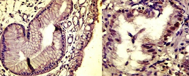
شملت الدراسة خمسين كتلة بارافينية من مرضى مصابين بأورام المعدة الحميدة (17 ذكراً، 33 أنثى). صنفت 44 حالة على أنها أورام حميدة زائدة، و3 حالات كأورام حميدة زائدة مع خلايا غير طبيعية، و3 حالات كأورام غدية. أظهرت معظم الأورام الحميدة الزائدة بالمعدة صبغا بدرجات مختلفة. أظهرت جميع الأورام الحميدة الزائدة بالمعدة ذات الخلايا غير الطبيعية صبغا نوويا؛ بينما أظهر اثنان من أصل ثلاثة أورام غدية بالمعدة صبغا غائبا.
الاستنتاج
لم يكن هناك ارتباط معنوي بين تعبير بروتين P53 ونوع أورام المعدة الحميدة، أو وجود التحول المعوي، أو بكتيريا هيليكوباكتر بيلوري. ومع ذلك، كان هناك ارتباط معنوي مع حجم الأورام. تحتاج نتائجنا إلى تأكيد من خلال دراسات أخرى لتحديد فائدة P53 كعلامة تشخيصية.
التنزيلات
المراجع
Hu H, Zhang Q, Chen G, Pritchard DM, Zhang S. Risk factors and clinical correlates of neoplastic transformation in gastric hyperplastic polyps in Chinese patients. Sci Rep. 2020;10(1). doi: 10.1038/s41598-020-58900-z. DOI: https://doi.org/10.1038/s41598-020-58900-z
João M, Areia M, Alves S, Elvas L, Taveira F, Brito D, et al. Gastric hyperplastic polyps: A benign entity? Analysis of recurrence and neoplastic transformation in a cohort study. Portuguese J Gastroenterol. 2021;28(5):328–335. doi: 10.1159/000514714. DOI: https://doi.org/10.1159/000514714
Yantiss RK. Immunohistochemical and molecular features of gastric hyperplastic polyps. Adv Cytol Pathol. 2017;2(1). doi: 10.15406/acp.2017.02.00012. DOI: https://doi.org/10.15406/acp.2017.02.00012
Sionov RV, Haupt Y. The cellular response to p53: the decision between life and death. Oncogene. 1999;18(45):6145–6157. doi: 10.1038/sj.onc.1203130. DOI: https://doi.org/10.1038/sj.onc.1203130
Prives C, Hall PA. The p53 pathway. J Pathol. 1999;187(1):112–126. PMID: 10341712. DOI: https://doi.org/10.1002/(SICI)1096-9896(199901)187:1<112::AID-PATH250>3.3.CO;2-V
Vousden KH, Lu X. Live or let die: the cell's response to p53. Nat Rev Cancer. 2002;2(8):594-604. doi: 10.1038/nrc864. DOI: https://doi.org/10.1038/nrc864
Lacroix M, Toillon RA, Leclercq G. p53 and breast cancer, an update. Endocr Relat Cancer. 2006;13(2):293–325. doi: 10.1677/erc.1.01172. DOI: https://doi.org/10.1677/erc.1.01172
Kubbutat MHG, Jones SN, Vousden KH. Regulation of p53 stability by Mdm2. Nature. 1997;387(6630):299–303. doi: 10.1038/387299a0. DOI: https://doi.org/10.1038/387299a0
Markowski AR, Markowska A, Guzinska-Ustymowicz K. Pathophysiological and clinical aspects of gastric hyperplastic polyps. World J Gastroenterol. 2016;22(40):8883. doi: 10.3748/wjg.v22.i40.8883. DOI: https://doi.org/10.3748/wjg.v22.i40.8883
Zhang X, Wang M, Wang Y, Cheng X, Jiang Y, Xiao H. Clinicopathologic significance of Her-2 and P53 expressions in gastric cancer. Asian J Surg. 2023;46(1):526–531. doi: 10.1016/j.asjsur.2022.06.039. DOI: https://doi.org/10.1016/j.asjsur.2022.06.039
Kumar V, Abbas AK, Aster JC, (Eds.), Robbins Basic Pathology, (10th Ed.), Elsevier - Health Sciences Division, 2017.
Yamanaka K, Miyatani H, Yoshida Y, Ishii T, Asabe S, Takada O, et al. Malignant transformation of a gastric hyperplastic polyp in a context of Helicobacter pylori-negative autoimmune gastritis: a case report. BMC Gastroenterol. 2016;16(1). doi: 10.1186/s12876-016-0537-x. DOI: https://doi.org/10.1186/s12876-016-0537-x
Dascălu RI, Păduraru DN, Bolocan A, Ion D, Andronic O. The role of PET-CT in gastric cancer – A narrative review. Sudan J Med Sci. 2020;332–44332–44344. doi: 10.18502/sjms.v15i3.7749. DOI: https://doi.org/10.18502/sjms.v15i3.7749
Ahn JY, Son DH, Choi KD, Roh J, Lim H, Choi KS, et al. Neoplasms arising in large gastric hyperplastic polyps: endoscopic and pathologic features. Gastrointest Endosc. 2014;80(6):1005-1013. doi: 10.1016/j.gie.2014.04.020. DOI: https://doi.org/10.1016/j.gie.2014.04.020
Terada T. Malignant transformation of foveolar hyperplastic polyp of the stomach: a histopathological study. Med Oncol. 2010;28(4):941–944. doi: 10.1007/s12032-010-9556-6. DOI: https://doi.org/10.1007/s12032-010-9556-6
Yao T, Kajiwara M, Kuroiwa S, Iwashita A, Oya M, Kabashima A, et al. Malignant transformation of gastric hyperplastic polyps: Alteration of phenotypes, proliferative activity, and p53 expression. Hum Pathol. 2002;33(10):1016–1022. doi: 10.1053/hupa.2002.126874. DOI: https://doi.org/10.1053/hupa.2002.126874
Imura J, Hayashi S, Ichikawa K, Miwa S, Nakajima T, Nomoto K, et al. Malignant transformation of hyperplastic gastric polyps: An immunohistochemical and pathological study of the changes of neoplastic phenotype. Oncol Lett. 2014;7(5):1459–463. doi: 10.3892/ol.2014.1932. DOI: https://doi.org/10.3892/ol.2014.1932
Murakami K, Mitomi H, Yamashita K, Tanabe S, Saigenji K, Okayasu I. p53, but not c-Ki-ras, mutation and down-regulation of p21WAF1/CIP1and cyclin D1 are associated with malignant transformation in gastric hyperplastic polyps. Am J Clin Pathol. 2001;115(2):224–234. doi: 10.1309/VLF5-UCNH-XQM2-X410. DOI: https://doi.org/10.1309/VLF5-UCNH-XQM2-X410
التنزيلات
منشور
كيفية الاقتباس
إصدار
القسم
الرخصة
الحقوق الفكرية (c) 2024 Al-Rafidain Journal of Medical Sciences

هذا العمل مرخص بموجب Creative Commons Attribution-NonCommercial-ShareAlike 4.0 International License.
Published by Al-Rafidain University College. This is an open access journal issued under the CC BY-NC-SA 4.0 license (https://creativecommons.org/licenses/by-nc-sa/4.0/).