Non-Invasive Treatment of Reversible Caries Lesions in Vitro: A Novel Era in Denal Practice
DOI:
https://doi.org/10.54133/ajms.v8i1.1709Keywords:
CPP-ACP, Confocal Raman microscopy, Full-width at half-maximum, Non-invasive treatment, nHA containing-dentifrice, Phosphate mapsAbstract
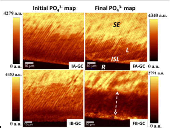
Background: The efficacy of GC Tooth Mousse cream (CPP-ACP) as a remineralizing agent has been affirmed. Recently, nano-hydroxyapatite-containing dentifrice “KAREX” has been put on the market as a dental care product suitable for dental tissue renovation. Objective: Using an in vitro caries model to compare the remineralizing effect of the two products. Methods: 12 sound premolars were exposed to pH cycling model to induce subsurface lesions. Thereafter, teeth were randomly divided into 2 groups scanned by Raman microscopy two times: once after initial caries induction and once again after intervention to provide phosphate maps showing the net differences between affected and unaffected enamel areas. Each specimen was treated with the respective remineralizing agent for 5 min every 24 h for 21 days. The prominent phosphate peak at 960 cm–1 was nominated to observe changes in its intensity. Results: The width of the phosphate peak measured by FWHM was calculated across each spectral map to evaluate the effect of remineralizing agents on the crystalline structure of demineralized enamel. At the end of the treatment, a significant difference has been attained in respect to phosphate gain in the body of lesions treated by nHA-containing dentifrice compared to the counterpart treated by CPP-ACP. However, no significant differences were observed among the treatment groups with regard to enamel crystallinity. Conclusions: Enamel surface layer permeability along with material consistency might represent key factors in subsurface lesion remineralization.
Downloads
References
Robinson C, Shore RC, Brookes SJ, Strafford S, Wood SR, Kirkham J. The chemistry of enamel caries. Crit Rev Oral Biol Med. 2000;11(4):481-495. doi: 10.1177/10454411000110040601.
Cross KJ, Huq NL, Reynolds EC. Casein phosphopeptides in oral health--chemistry and clinical applications. Curr Pharm Des. 2007;13(8):793-800. doi: 10.2174/138161207780363086. DOI: https://doi.org/10.2174/138161207780363086
Reynolds EC. Anticariogenic complexes of amorphous calcium phosphate stabilized by casein phosphopeptides: a review. Spec Care Dentist. 1998;18(1):8-16. doi: 10.1111/j.1754-4505.1998.tb01353.x. DOI: https://doi.org/10.1111/j.1754-4505.1998.tb01353.x
Reynolds EC, Johnson IH. Effect of milk on caries incidence and bacterial composition of dental plaque in the rat. Arch Oral Biol. 1981;26(5):445-451. doi: 10.1016/0003-9969(81)90042-x. DOI: https://doi.org/10.1016/0003-9969(81)90042-X
Reynolds EC, del Rio A. Effect of casein and whey-protein solutions on caries experience and feeding patterns of the rat. Arch Oral Biol. 1984;29(11):927-933. doi: 10.1016/0003-9969(84)90093-1. DOI: https://doi.org/10.1016/0003-9969(84)90093-1
Reynolds EC. Remineralization of enamel subsurface lesions by casein phosphopeptide-stabilized calcium phosphate solutions. J Dent Res. 1997;76(9):1587-1595. doi: 10.1177/00220345970760091101. DOI: https://doi.org/10.1177/00220345970760091101
Chen H, Liu X, Dai J, Jiang Z, Guo T, Ding Y. Effect of remineralizing agents on white spot lesions after orthodontic treatment: a systematic review. Am J Orthod Dentofacial Orthop. 2013;143(3):376-382. doi: 10.1016/j.ajodo.2012.10.013. DOI: https://doi.org/10.1016/j.ajodo.2012.10.013
Vandiver J, Dean D, Patel N, Bonfield W, Ortiz C. Nanoscale variation in surface charge of synthetic hydroxyapatite detected by chemically and spatially specific high-resolution force spectroscopy. Biomaterials. 2005;26(3):271-283. doi: 10.1016/j.biomaterials.2004.02.053. DOI: https://doi.org/10.1016/j.biomaterials.2004.02.053
Yamagishi K, Onuma K, Suzuki T, Okada F, Tagami J, Otsuki M, et al. Materials chemistry: a synthetic enamel for rapid tooth repair. Nature. 2005;433(7028):819. doi: 10.1038/433819a. DOI: https://doi.org/10.1038/433819a
Huang SB, Gao SS, Yu HY. Effect of nano-hydroxyapatite concentration on remineralization of initial enamel lesion in vitro. Biomed Mater. 2009;4(3):034104. doi: 10.1088/1748-6041/4/3/034104. DOI: https://doi.org/10.1088/1748-6041/4/3/034104
Roveri N, Battistella E, Bianchi CL, Foltran I, Foresti E, Iafisco M, et al. Surface enamel remineralization: biomimetic apatite nanocrystals and fluoride ions different effects. J Nanomater. 2009;2009:8. doi: 10.1155/2009/746383. DOI: https://doi.org/10.1155/2009/746383
Li L, Pan H, Tao J, Xu X, Mao C, Gu X, et al. Repair of enamel by using hydroxyapatite nanoparticles as the building blocks. J Mater Chem. 2008;18(34):4079-4084. doi: 10.1039/B806090H. DOI: https://doi.org/10.1039/b806090h
Kani T, Kani M, Isozaki A, Shintani H, Ohashi T, Tokumoto T. Effect to apatite-containing dentifrices on dental caries in school children. J Dent Health. 1989;39(1):104-109. DOI: https://doi.org/10.5834/jdh.39.104
Najibfard K, Ramalingam K, Chedjieu I, Amaechi BT. Remineralization of early caries by a nano-hydroxyapatite dentifrice. J Clin Dent. 2011;22(5):139-143. PMID: 22403978.
Al-Obaidi R, Salehi H, Desoutter A, Bonnet L, Etienne P, Terrer E, et al. Chemical & nano-mechanical study of artificial human enamel subsurface lesions. Sci Rep. 2018;8(1):4047. doi: 10.1038/s41598-018-22459-7. DOI: https://doi.org/10.1038/s41598-018-22459-7
Mehta R, Nandlal B, Prashanth S. Comparative evaluation of remineralization potential of casein phosphopeptide-amorphous calcium phosphate and casein phosphopeptide-amorphous calcium phosphate fluoride on artificial enamel white spot lesion: an in vitro light fluorescence study. Indian J Dent Res. 2013;24(6):681-689. doi: 10.4103/0970-9290.127610. DOI: https://doi.org/10.4103/0970-9290.127610
Sato Y, Sato T, Niwa M, Aoki H. Precipitation of octacalcium phosphates on artificial enamel in artificial saliva. J Mater Sci Mater Med. 2006;17(11):1173-1177. doi: 10.1007/s10856-006-0545-4. DOI: https://doi.org/10.1007/s10856-006-0545-4
Xu C, Reed R, Gorski JP, Wang Y, Walker MP. The distribution of carbonate in enamel and its correlation with structure and mechanical properties. J Mater Sci. 2012;47(23):8035-8043. doi: 10.1007/s10853-012-6693-7. DOI: https://doi.org/10.1007/s10853-012-6693-7
Featherstone JD, ten Cate JM, Shariati M, Arends J. Comparison of artificial caries-like lesions by quantitative microradiography and microhardness profiles. Caries Res. 1983;17(5):385-391. doi: 10.1159/000260692. DOI: https://doi.org/10.1159/000260692
Cross KJ, Huq NL, Palamara JE, Perich JW, Reynolds EC. Physicochemical characterization of casein phosphopeptide-amorphous calcium phosphate nanocomplexes. J Biol Chem. 2005;280(15):15362-15369. doi: 10.1074/jbc.M413504200. DOI: https://doi.org/10.1074/jbc.M413504200
Cochrane NJ, Saranathan S, Cai F, Cross KJ, Reynolds EC. Enamel subsurface lesion remineralisation with casein phosphopeptide stabilised solutions of calcium, phosphate and fluoride. Caries Res. 2008;42(2):88-97. doi: 10.1159/000113161. DOI: https://doi.org/10.1159/000113161
Cochrane N, Reynolds E. Casein phosphopeptides in oral health. Food constituents and oral health: Elsevier; 2009. p. 185-224. DOI: https://doi.org/10.1533/9781845696290.2.185
Huq NL, Cross KJ, Reynolds EC. A 1H-NMR study of the casein phosphopeptide alpha s1-casein(59-79). Biochim Biophys Acta. 1995;1247(2):201-208. PMID: 7696309. DOI: https://doi.org/10.1016/0167-4838(94)00229-A
Cross KJ, Huq NL, O’Brien-Simpson NM, Perich JW, Attard TJ, Reynolds EC. The role of multiphosphorylated peptides in mineralized tissue regeneration. Int J Peptide Res Ther. 2007;13(4):479-495. DOI: https://doi.org/10.1007/s10989-007-9105-0
Larsen MJ, Jensen SJ. Solubility study of the initial formation of calcium orthophosphates from aqueous solutions at pH 5-10. Arch Oral Biol. 1986;31(9):565-572. doi: 10.1016/0003-9969(86)90079-8. DOI: https://doi.org/10.1016/0003-9969(86)90079-8
Larsen MJ, Fejerskov O. Chemical and structural challenges in remineralization of dental enamel lesions. Scand J Dent Res. 1989;97(4):285-296. doi: 10.1111/j.1600-0722.1989.tb01615.x.
Akkus A, Akkus A, Roperto R, Akkus O, Porto T, Teich S, et al. Evaluation of mineral content in healthy permanent human enamel by Raman spectroscopy. J Clin Exp Dent. 2016;8(5):e546-e549. doi: 10.4317/jced.53057. DOI: https://doi.org/10.4317/jced.53057
ten Cate JM, Arends J. Remineralization of artificial enamel lesions in vitro: III. A study of the deposition mechanism. Caries Res. 1980;14(6):351-358. doi: 10.1159/000260477. DOI: https://doi.org/10.1159/000260477
Silverstone LM, Wefel JS, Zimmerman BF, Clarkson BH, Featherstone MJ. Remineralization of natural and artificial lesions in human dental enamel in vitro. Effect of calcium concentration of the calcifying fluid. Caries Res. 1981;15(2):138-157. doi: 10.1159/000260512. DOI: https://doi.org/10.1159/000260512
Larsen MJ, Fejerskov O. Chemical and structural challenges in remineralization of dental enamel lesions. Scand J Dent Res. 1989;97(4):285-296. doi: 10.1111/j.1600-0722.1989.tb01615.x. DOI: https://doi.org/10.1111/j.1600-0722.1989.tb01615.x
Larsen MJ. An investigation of the theoretical background for the stability of the calcium-phosphate salts and their mutual conversion in aqueous solutions. Arch Oral Biol. 1986;31(11):757-761. doi: 10.1016/0003-9969(86)90008-7. DOI: https://doi.org/10.1016/0003-9969(86)90008-7
Andersson A, Sköld-Larsson K, Hallgren A, Petersson LG, Twetman S. Effect of a dental cream containing amorphous cream phosphate complexes on white spot lesion regression assessed by laser fluorescence. Oral Health Prev Dent. 2007;5(3):229-233. PMID: 17977295.
Bailey DL, Adams GG, Tsao CE, Hyslop A, Escobar K, Manton DJ, et al. Regression of post-orthodontic lesions by a remineralizing cream. J Dent Res. 2009;88(12):1148-1153. doi: 10.1177/0022034509347168. DOI: https://doi.org/10.1177/0022034509347168
Beerens MW, van der Veen MH, van Beek H, ten Cate JM. Effects of casein phosphopeptide amorphous calcium fluoride phosphate paste on white spot lesions and dental plaque after orthodontic treatment: a 3-month follow-up. Eur J Oral Sci. 2010;118(6):610-617. doi: 10.1111/j.1600-0722.2010.00780.x. DOI: https://doi.org/10.1111/j.1600-0722.2010.00780.x
Bröchner A, Christensen C, Kristensen B, Tranæus S, Karlsson L, Sonnesen L, et al. Treatment of post-orthodontic white spot lesions with casein phosphopeptide-stabilised amorphous calcium phosphate. Clin Oral Investig. 2011;15(3):369-373. doi: 10.1007/s00784-010-0401-2. DOI: https://doi.org/10.1007/s00784-010-0401-2
Stylianopoulos T, Poh MZ, Insin N, Bawendi MG, Fukumura D, Munn LL, et al. Diffusion of particles in the extracellular matrix: the effect of repulsive electrostatic interactions. Biophys J. 2010;99(5):1342-1349. doi: 10.1016/j.bpj.2010.06.016. DOI: https://doi.org/10.1016/j.bpj.2010.06.016
Kaehler T. Nanotechnology: basic concepts and definitions. Clin Chem. 1994 Sep;40(9):1797-1799. PMID: 8070103. DOI: https://doi.org/10.1093/clinchem/40.9.1797b
ten Cate JM, Jongebloed WL, Arends J. Remineralization of artificial enamel lesions in vitro. IV. Influence of fluorides and diphosphonates on short- and long-term reimineralization. Caries Res. 1981;15(1):60-69. doi: 10.1159/000260501. DOI: https://doi.org/10.1159/000260501
Weatherell JA, Robinson C, Hiller CR. Distribution of carbonate in thin sections of dental enamel. Caries Res. 1968;2(1):1-9. doi: 10.1159/000259538. DOI: https://doi.org/10.1159/000259538
Robinson C, Weatherell JA, Hallsworth AS. Variatoon in composition of dental enamel within thin ground tooth sections. Caries Res. 1971;5(1):44-57. doi: 10.1159/000259731. DOI: https://doi.org/10.1159/000259731
Robinson C, Shore RC, Brookes SJ, Strafford S, Wood SR, Kirkham J. The chemistry of enamel caries. Cri Rev Oral Biol Med. 2000;11(4):481-495. doi:10.1177/10454411000110040601. DOI: https://doi.org/10.1177/10454411000110040601
Downloads
Published
How to Cite
Issue
Section
License
Copyright (c) 2025 Al-Rafidain Journal of Medical Sciences ( ISSN 2789-3219 )

This work is licensed under a Creative Commons Attribution-NonCommercial-ShareAlike 4.0 International License.
Published by Al-Rafidain University College. This is an open access journal issued under the CC BY-NC-SA 4.0 license (https://creativecommons.org/licenses/by-nc-sa/4.0/).


