Comparison Between Impacted Maxillary and Mandibular Canines in an Iraqi Population: Prevalence and Classification
DOI:
https://doi.org/10.54133/ajms.v8i1.1690Keywords:
CBCT, Impacted canine, Prevalence classificationAbstract
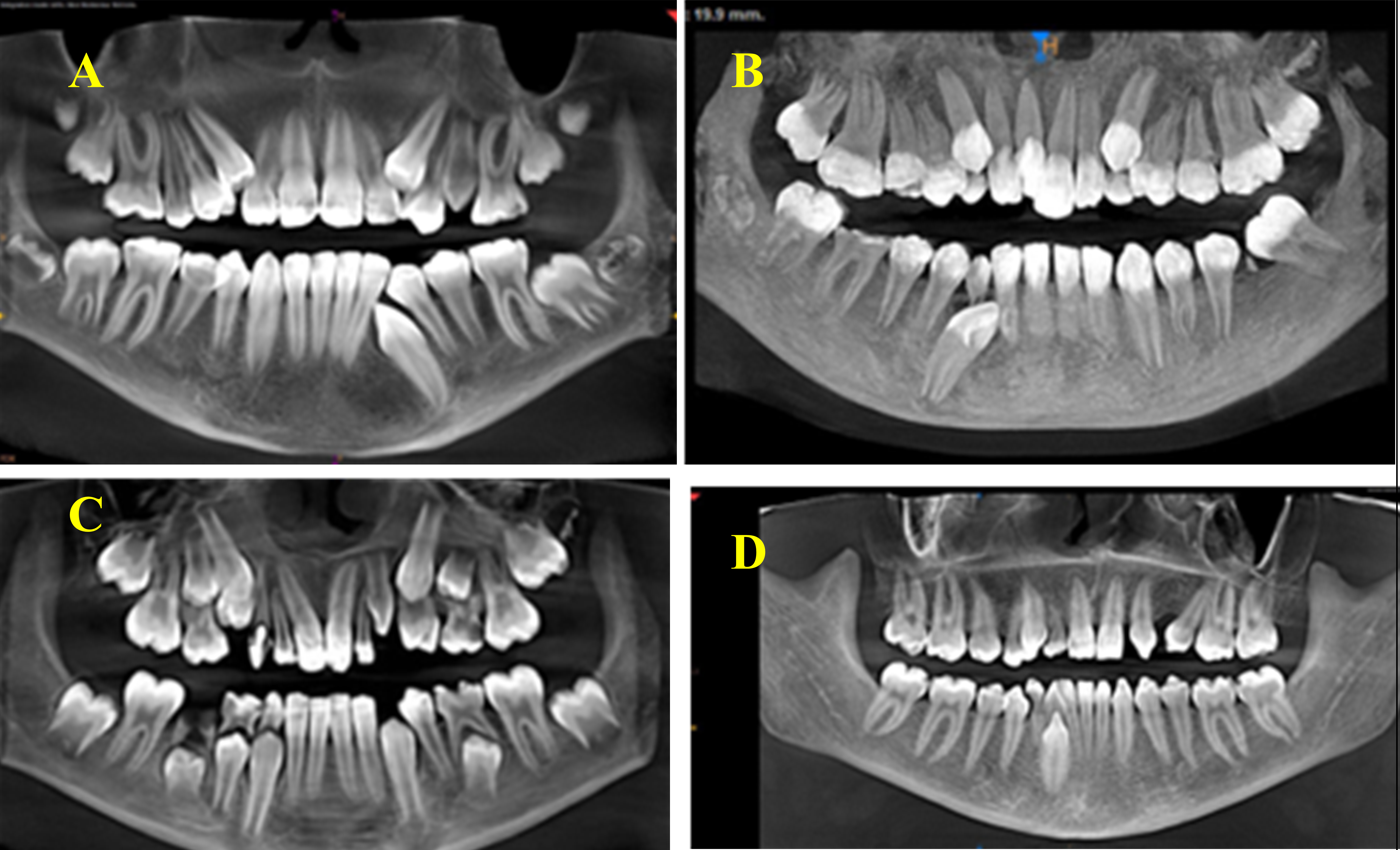
Background: Concerning the importance of impacted canines for aesthetics and function to improve patients’ health, it is crucial to provide the oral surgeon and orthodontist with a complete analysis of their location, angulation, and relation with adjacent teeth. Objective: To determine and compare the frequencies of different types of impacted maxillary and mandibular canines under the current classification systems in an Iraqi population sample. Methods: This study retrospectively examined the cone beam computed tomography scans of 1000 Iraqi patients aged 12–40 years (380 males and 620 females) who had attended the Oral and Maxillofacial Radiology Department at Ghazi Al-Hariri and Al-Sadder City Hospitals. Results: Of the 1000 patients, 49 had impacted maxillary canines (4.9%), of which 18(36.7%) were male and 31(63.3%) were female. Bilateral impaction was more common than unilateral impaction (61.2% vs. 19.0%). Type II was the most common impaction type. In addition, 20 patients had impacted mandibular canines (2%), of which 8(40.0%) were male and 12(60.0%) were female. Bilateral impaction was less common than unilateral impaction (25.0% vs. 75.0%). Type III was the most common impaction. Conclusions: Impaction was more common for maxillary canines (4.9%) than for mandibular canines (2.0%). Type II impaction was the most common for maxillary canines, followed by Types I, IV, and VII. In contrast, Type III impaction was the most common for mandibular canines, followed by Type V.
Downloads
References
Bass TB. Observations on the misplaced upper canine tooth. Dent Pract Dent Rec. 1967;18(1):25-33. PMID: 5235604.
Pedro FL, Bandéca MC, Volpato LE, Marques AT, Borba AM, Musis CR, et al. Prevalence of impacted teeth in a Brazilian subpopulation. J Contemp Dent Pract. 2014;15(2):209-213. doi: 10.5005/jp-journals-10024-1516. DOI: https://doi.org/10.5005/jp-journals-10024-1516
Chung DD, Weisberg M, Pagala M. Incidence and effects of genetic factors on canine impaction in an isolated Jewish population. Am J Orthod Dentofacial Orthop. 2011;139: 331-335. doi: 10.1016/j.ajodo.2010.06.023. DOI: https://doi.org/10.1016/j.ajodo.2010.06.023
Sajnani AK, King NM. Impacted mandibular canines: prevalence and characteristic features in southern Chinese children and adolescents. J Dent Child (Chic). 2014;81(1):3-6. PMID: 24709426.
Yavuz MS, Aras MH, Büyükkurt MC, Tozoglu S. Impacted mandibular canines. J Contemp Dent Pract. 2007;8(7):78-85. PMID: 17994158. DOI: https://doi.org/10.5005/jcdp-8-7-78
Mason C, Papadakou P, Roberts GJ. The radiographic localization of impacted maxillary canines: a comparison of methods. Eur J Orthod. 2001;23(1):25-34. doi: 10.1093/ejo/23.1.25. DOI: https://doi.org/10.1093/ejo/23.1.25
Kuftinec MM, Shapira Y. The impacted maxillary canine: I. Review of concepts. ASDC J Dent Child. 1995;62(5):317-324. PMID: 8550920.
Hudson AP, Harris AM, Mohamed N. Early identification and management of mandibular canine ectopia. SADJ. 2011;66(10):462-464, PMID: 23193881.
Celikoglu M, Kamak H, Oktay H. Investigation of transmigrated and impacted maxillary and mandibular canine teeth in an orthodontic patient population. J Oral Maxillofac Surg. 2010;68(5):1001-1006. doi: 10.1016/j.joms.2009.09.006. DOI: https://doi.org/10.1016/j.joms.2009.09.006
Aktan AM, Kara S, Akgünlü F, Malkoç S. The incidence of canine transmigration and tooth impaction in a Turkish subpopulation. Eur J Orthod. 2010;32(5):575-581. doi: 10.1093/ejo/cjp151. DOI: https://doi.org/10.1093/ejo/cjp151
Richardson G, Russell KA. A review of impacted permanent maxillary cuspids--diagnosis and prevention. J Can Dent Assoc. 2000;66(9):497-501. PMID: 11070629.
Jung YH, Liang H, Benson BW, Flint DJ, Cho BH. The assessment of impacted maxillary canine position with panoramic radiography and cone beam CT. Dentomaxillofac Radiol. 2012;41(5):356-360. doi: 10.1259/dmfr/14055036. DOI: https://doi.org/10.1259/dmfr/14055036
Alrahabi M, Sohail Zafar M. Evaluation of root canal morphology of maxillary molars using cone beam computed tomography. Pak J Med Sci. 2015;31(2):426-430. doi: 10.12669/pjms.312.6753. DOI: https://doi.org/10.12669/pjms.312.6753
Noaman AT, Bede SY. The effect of bone density measured by cone beam computed tomography and implant dimensions on the stability of dental implants. J Craniofac Surg. 2022;33(6):e553-e557. doi: 10.1097/SCS.0000000000008429. DOI: https://doi.org/10.1097/SCS.0000000000008429
Lai CS, Bornstein MM, Mock L, Heuberger BM, Dietrich T, Katsaros C. Impacted maxillary canines and root resorptions of neighbouring teeth: a radiographic analysis using cone-beam computed tomography. Eur J Orthod. 2013;35(4):529-538. doi: 10.1093/ejo/cjs037. DOI: https://doi.org/10.1093/ejo/cjs037
Rahman VF, Fatah AA. Localization of maxillary impacted canine using cone beam computed tomography for assessment of angulation, distance from occlusal Plane, alveolar width and proximity to adjacent teeth. J Baghdad Coll Dent. 2017; 29(1):70-75. DOI: https://doi.org/10.12816/0038627
Al-Khawaja NFK, Nahidh M, Abdulsaheb RJ. Assessment of maxillary incisors' angulation and position in different types of malocclusions using cone-beam computed tomography. Contemp Clin Dent. 2021;12(4):401-407. doi: 10.4103/ccd.ccd_743_20. DOI: https://doi.org/10.4103/ccd.ccd_743_20
Yamamoto G, Ohta Y, Tsuda,Y, Tanaka A, Nishikawa M, Inoda H. A new classification of impacted canines and second premolars using orthopantomography. Asian J Oral Maxillofacl Surg. 2003;15(1):31-37. doi: 10.1016/S0915-6992(03)80029-8. DOI: https://doi.org/10.1016/S0915-6992(03)80029-8
Mupparapu M. Patterns of intra-osseous transmigration and ectopic eruption of mandibular canines: review of literature and report of nine additional cases. Dentomaxillofac Radiol. 2002;31(6):355-360. doi: 10.1038/sj.dmfr.4600732. DOI: https://doi.org/10.1038/sj.dmfr.4600732
Rózsa N, Fábián G, Szádeczky B, Kaán M, Gábris K, Tarján I. Prevalence of impacted permanent upper canine and its treatment in 11-18-year-old orthodontic patients. Fogorv Sz. 2003;96(2):65-69. PMID: 12762148.
Andreasen JO, Peterson J, Laskin DM, (Eds.), Textbook and Color Atlas of Tooth Impactions—Diagnosis, Treatment and Prevention. Mosby Year Book, St Louis. 1997.
Grover PS, Lorton L. The incidence of unerupted permanent teeth and related clinical cases. Oral Surg Oral Med Oral Pathol. 1985;59:420-425. doi: 10.1016/0030-4220(85)90070-2. DOI: https://doi.org/10.1016/0030-4220(85)90070-2
Chu FC, Li TK, Lui VK, Newsome PR, Chow RL, Cheung LK. Prevalence of impacted teeth and associated pathologies--a radiographic study of the Hong Kong Chinese population. Hong Kong Med J. 2003;9(3):158-163. PMID: 12777649.
Lazim AI. The prevalence of impacted maxillary canine among Iraqi patients of Al-Basrah City. J Baghdad Coll Dent. 2017; 28(1):73-77 DOI: https://doi.org/10.12816/0024712
Sajnani AK, King NM. Impacted mandibular canines: prevalence and characteristic features in southern Chinese children and adolescents. J Dent Child (Chic). 2014 ;81(1):3-6. PMID: 24709426.
Aydin U, Yilmaz HH, Yildirim D. Incidence of canine impaction and transmigration in a patient population. Dentomaxillofac Radiol. 2004;33(3):164-169. doi: 10.1259/dmfr/15470658. DOI: https://doi.org/10.1259/dmfr/15470658
Topkara A, Sari Z. Impacted teeth in a Turkish orthodontic patient population: prevalence, distribution and relationship with dental arch characteristics. Eur J Paediatr Dent. 2012;13(4):311-316. PMID: 23270290.
Agastra E, Saettone M, Parrini S, Cugliari G, Deregibus A, Castroflorio T. Impacted permanent mandibular canines: Epidemiological evaluation. J Clin Med. 2023;12(16):5375. doi: 10.3390/jcm12165375. DOI: https://doi.org/10.3390/jcm12165375
Sanu OO, Adeyemi TA, Isiekwe MC. Incidence of impacted mandibular canine and associated pathologies in an orthodontic patient population in Lagos, Nigeria. Nig Q J Hosp Med. 2012;22(4):291-295. PMID: 24596969.
Al-Zoubi H, Alharbi AA, Ferguson DJ, Zafar MS. Frequency of impacted teeth and categorization of impacted canines: A retrospective radiographic study using orthopantomograms. Eur J Dent. 2017;11(1):117-121. doi: 10.4103/ejd.ejd_308_16. DOI: https://doi.org/10.4103/ejd.ejd_308_16
Gashi A, Kamberi B, Ademi-Abdyli R, Perjuci F, Sahatçiu-Gashi A. The incidence of impacted maxillary canines in a Kosovar Population. Int Sch Res Notices. 2014;2014:370531. doi: 10.1155/2014/370531. DOI: https://doi.org/10.1155/2014/370531
Al Fawzan AA, Alruwaithi M, Alsadoon S. Prevalence of maxillary canine impaction in orthodontics at Eastern Riyadh specialized dental center. IOSR J Dent Med Sci. 2017;16(1):72-74. doi: 10.9790/0853-1601057274. DOI: https://doi.org/10.9790/0853-1601057274
Downloads
Published
How to Cite
Issue
Section
License
Copyright (c) 2025 Al-Rafidain Journal of Medical Sciences ( ISSN 2789-3219 )

This work is licensed under a Creative Commons Attribution-NonCommercial-ShareAlike 4.0 International License.
Published by Al-Rafidain University College. This is an open access journal issued under the CC BY-NC-SA 4.0 license (https://creativecommons.org/licenses/by-nc-sa/4.0/).


