Apparent Diffusion Coefficient in the Diagnosis and Follow-up of Multiple Sclerosis: Role of Magnetic Resonance Imaging
DOI:
https://doi.org/10.54133/ajms.v7i1.1114Keywords:
Apparent diffusion coefficient (ADC), Brain MRI, Multiple sclerosisAbstract
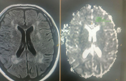
Background: Chronic Multiple Sclerosis (MS) modifies the apparent diffusion coefficient (ADC) value due to severe pathological changes. After trauma, it is the second most common cause of brain injury in healthy young adults. MRI is considered the initial imaging modality for MS diagnosis and follow-up. Objective: To assess the significance of the ADC in the diagnosis and follow-up of MS plaques across various disease subtypes. Methods: Forty MS patients were included in a case-control study at Ibn-Sina Teaching Hospital, Mosul Province, between June 1, 2022 and February 28, 2023. The patients had diffusion-weighted and traditional MR imaging with ADC measurement in plaques, and the normal white matter value of controls was compared to the patients' results. Results: The ADC values were higher in cases that were acute or secondary-progressive than in relapsing-remitting cases or normal white matter. In both types of newly generated plaques, there was an initial non-significant increase in ADC values compared to existing plaques. Overall, the ADC sensitivity, specificity, and accuracy in diagnosing MS were 85.7%, 95.2%, and 90.5% in acute cases, and 85.7%, 83.3%, and 84.6% in chronic cases, respectively, with no significant difference between active and inactive lesions. Conclusions: The apparent diffusion coefficient value can be included in the imaging protocol for the diagnosis and follow-up of various subtypes of MS.
Downloads
References
Confavreux C, Vukusic S, Moreau T, Adeleine P. Relapses and progression of disability in multiple sclerosis. N Engl J Med. 2000;343(20):1430-1438. doi: 10.1056/NEJM200011163432001. DOI: https://doi.org/10.1056/NEJM200011163432001
Sarbu N, Shih RY, Jones RV, Horkayne-Szakaly I, Oleaga L, Smirniotopoulos JG. White matter diseases with radiologic-pathologic correlation. Radiographics. 2016;36(5):1426-1447. doi: 10.1148/rg.2016160031. DOI: https://doi.org/10.1148/rg.2016160031
Okuda DT, Mowry EM, Beheshtian A, Waubant E, Baranzini SE, Goodin DS, et al. Incidental MRI anomalies suggestive of multiple sclerosis: the radiologically isolated syndrome. Neurology. 2009;72(9):800-805. doi: 10.1212/01.wnl.0000335764.14513.1a. DOI: https://doi.org/10.1212/01.wnl.0000335764.14513.1a
Garaci FG, Marziali S, Meschini A, Fornari M, Rossi S, Melis M, et al. Brain hemodynamic changes associated with chronic cerebrospinal venous insufficiency are not specific to multiple sclerosis and do not increase its severity. Radiology. 2012;265(1):233-239. doi: 10.1148/radiol.12112245. DOI: https://doi.org/10.1148/radiol.12112245
Bakshi R, Minagar A, Jaisani Z, Wolinsky JS. Imaging of multiple sclerosis: role in neurotherapeutics. NeuroRx. 2005;2(2):277-303. doi: 10.1602/neurorx.2.2.277. DOI: https://doi.org/10.1602/neurorx.2.2.277
Mohammed F, Ismail M. Validity of apparent diffusion coefficient value in diagnosis and follow up of multiple sclerosis patients in different clinical subtypes. Egypt J Radiol Nuclear Med. 2018;49(4):1103-1109. doi: 10.1016/j.ejrnm.2018.07.006. DOI: https://doi.org/10.1016/j.ejrnm.2018.07.006
McNamara C, Sugrue G, Murray B, MacMahon PJ. Current and emerging therapies in multiple sclerosis: Implications for the radiologist, Part 1-Mechanisms, efficacy, and safety. Am J Neuroradiol. 2017;38(9):1664-1671. doi: 10.3174/ajnr.A5147. DOI: https://doi.org/10.3174/ajnr.A5147
Daoud AG, Waheed HJ, Abdala ME. The Impact of Vitamin D on The Development of Multiple Sclerosis (Review article). Al Mustansiriyah J Pharm Sci. 2022;21(3):7–15. doi: 10.32947/ajps.v21i3.792. DOI: https://doi.org/10.32947/ajps.v21i3.792
Salih KM, Abdullah AH, Sheaheed NM. Assessment of some platelet activating markers and secretory status with clinical manifestations in multiple sclerosis Iraqi patients. Al-Mustansiriyah J Sci. 2022;33(3):12-19. doi:10.23851/mjs.v33i3.1130. DOI: https://doi.org/10.23851/mjs.v33i3.1130
Zabirova AK, Bakulin IS, Poydasheva AG, Zakharova MN, Suponeva NA. Cognitive impairment and its treatment in patients with multiple sclerosis. Almanac Clin Med. 2023;51(2):110-125. doi: 10.18786/2072-0505-2023-51-009. DOI: https://doi.org/10.18786/2072-0505-2023-51-009
Chang CA, Chong AL, Chandra RV, Butler E, Rajendran D, Chuah K, et al. Detection of multiple sclerosis lesions in the cervical cord: which of the MAGNIMS 'mandatory' non-gadolinium enhanced sagittal sequences is optimal at 3T? Neuroradiol J. 2021;34(6):600-606. doi: 10.1177/19714009211017787. DOI: https://doi.org/10.1177/19714009211017787
Thompson AJ, Banwell BL, Barkhof F, Carroll WM, Coetzee T, Comi G, et al. Diagnosis of multiple sclerosis: 2017 revisions of the McDonald criteria. Lancet Neurol. 2018;17(2):162-173. doi: 10.1016/S1474-4422(17)30470-2. DOI: https://doi.org/10.1016/S1474-4422(17)30470-2
Hartung HP, Graf J, Aktas O, Mares J, Barnett MH. Diagnosis of multiple sclerosis: revisions of the McDonald criteria 2017 - continuity and change. Curr Opin Neurol. 2019;32(3):327-337. doi: 10.1097/WCO.0000000000000699. DOI: https://doi.org/10.1097/WCO.0000000000000699
Fahad QA, Hadi AS, Obaid SF. A comparison of sagittal sections of short T1inversion recovery and T2 weighted fast spin echo magnetic resonance sequences for detection of multiple sclerosis spinal cord lesions. Al-Kindy Coll Med J. 2016;12(2):60-63.
Filippi M, Inglese M. Overview of diffusion-weighted magnetic resonance studies in multiple sclerosis. J Neurol Sci. 2001;186 (Suppl 1):S37-43. doi: 10.1016/s0022-510x(01)00489-0. DOI: https://doi.org/10.1016/S0022-510X(01)00489-0
Filippi M, Iannucci G, Cercignani M, Assunta Rocca M, Pratesi A, Comi G. A quantitative study of water diffusion in multiple sclerosis lesions and normal-appearing white matter using echo-planar imaging. Arch Neurol. 2000;57(7):1017–1021. doi:10.1001/archneur.57.7.1017. DOI: https://doi.org/10.1001/archneur.57.7.1017
Almolla RM, Hassan HA, Raya YM, Hussein RA. Correlation of apparent diffusion coefficient to cognitive impairment in relapsing remittent multiple sclerosis (plaque, peri-plaque and normal appearing white matter). Egypt J Radiol Nucl Med. 2016;47:1009-1018. doi: 10.1016/j.ejrnm.2016.04.018. DOI: https://doi.org/10.1016/j.ejrnm.2016.04.018
Castriota Scanderbeg A, Tomaiuolo F, Sabatini U, Nocentini U, Grasso MG, Caltagirone C. Demyelinating plaques in relapsingremitting and secondary-progressive multiple sclerosis: assessment with diffusion MR imaging. Am J Neuroradiol. 2000;21(5):862-868. PMID: 10815661.
Mohamed MM, Al-Ani MI, Al Gawwam G, Alrubaye MH, Al-Imam A. Detection of multiple sclerosis lesions in supra- and infra-tentorial anatomical regions by double inversion recovery, flair, and T2 MRI sequences: A comparative study in Iraqi patients. Al-Rafidain J Med Sci. 2023;5(Suppl 1):S172-176. doi: 10.54133/ajms.v5i1S.357. DOI: https://doi.org/10.54133/ajms.v5i1S.357
Ragheb S, Ekladious M. The value of apparent diffusion coefficient measurement in assessment and follow up of multiple sclerosis patients. Egypt J Hospital Med. 2021;82(4):655-662. doi: 10.21608/EJHM.2021.150438. DOI: https://doi.org/10.21608/ejhm.2021.150438
Davoudi Y, Foroughipour M, Torabi R, Layegh P, Matin N, Shoeibi A. Diffusion weighted imaging in acute attacks of multiple sclerosis. Iran J Radiol. 2016;13(2):e21740. doi: 10.5812/iranjradiol.21740. DOI: https://doi.org/10.5812/iranjradiol.21740
Terzi M, Atalay K, Incesu L, Diren B, Onar M. Diffusion-weighted magnetic resonance imaging of the brain in multiple sclerosis. Turk J Neurol. 2007;13:310-318.
Unal S, Peker E, Erdogan S, Erden MI. Is it possible to discriminate active MS lesions with diffusion weighted imaging? Eurasian J Med. 2019;51(3):219-223. doi: 10.5152/eurasianjmed.2019.18473. DOI: https://doi.org/10.5152/eurasianjmed.2019.18473
Downloads
Published
How to Cite
Issue
Section
License
Copyright (c) 2024 Al-Rafidain Journal of Medical Sciences ( ISSN 2789-3219 )

This work is licensed under a Creative Commons Attribution-NonCommercial-ShareAlike 4.0 International License.
Published by Al-Rafidain University College. This is an open access journal issued under the CC BY-NC-SA 4.0 license (https://creativecommons.org/licenses/by-nc-sa/4.0/).


