Unusual Location for Common Tumor‒Vulvar Leiomyoma in a 40-Year-Old Woman: A Case Report and Review
DOI:
https://doi.org/10.54133/ajms.v9i2.2454Keywords:
Case report, Fibroid, Vulvar leiomyoma, Vulvar massAbstract
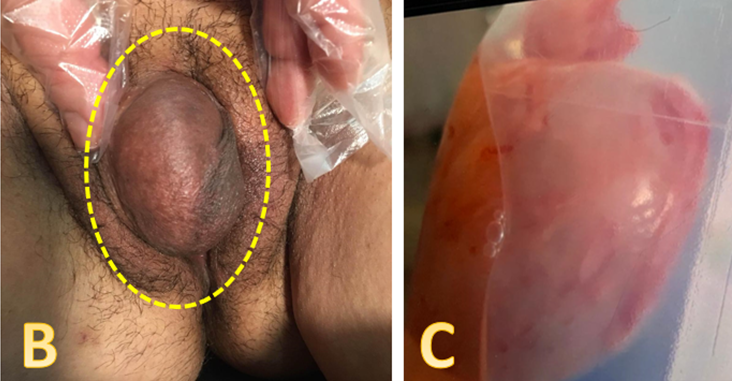
Leiomyomas are common benign smooth muscle tumors of the uterus; their presentation in the vulva is rare and tends to impose diagnostic challenges, mistaken for other conditions. A forty-year-old, perimenopausal, unmarried patient attended our clinic complaining of a slowly growing vulvar mass for the last 6 months. General evaluation and past medical history were unremarkable. Upon local examination, a firm, non-tender vulvar mass measuring 3 × 4 cm was found, causing discomfort during walking. Preoperative lab investigation and ultrasonography suggested a benign tumor. The patient was advised to undergo surgical excision, which was done under general anesthesia. Histopathology confirms the case. Postoperative recovery was uneventful; she was followed every 6 months; no recurrence after 5 years of follow-up. Vulvar leiomyomas, although rare, should be excluded in a vulvar mass. Definite diagnosis requires histopathological confirmation. Surgical excision of the whole mass provides effective treatment and a better prognosis.
Downloads
References
Sun C, Zou J, Wang Q, Wang Q, Han L, Batchu N, et al. Review of the pathophysiology, diagnosis, and therapy of vulvar leiomyoma, a rare gynecological tumor. J Int Med Res. 2018;46(2):663-674. doi: 10.1177/0300060517721796. DOI: https://doi.org/10.1177/0300060517721796
Goyal LD, Garg P, Kaur M. Vulvar leiomyoma in an adolescent girl: a case report and review of the literature. J Med Case Rep. 2023;17(1):68. doi: 10.1186/s13256-022-03743-7. DOI: https://doi.org/10.1186/s13256-022-03743-7
Hamada E, Shiga N, Watanabe M, Sato S, Watanabe Z, Tachibana M, et al. A rare case of uterine‐origin vulvar leiomyoma occurring in a juvenile girl: Case report and literature review. Case Rep Obstetr Gynecol. 2025;2025(1):8874924. doi: 10.1155/crog/8874924. DOI: https://doi.org/10.1155/crog/8874924
Baradwan S, Latifah HM, Al-Maghrabi H, Almutawa AM. Vulvar leiomyoma mimicking Bartholin's gland cyst: a case report. Case Rep Women's Health. 2024;44:e00655. doi: 10.1016/j.crwh.2024.e00655. DOI: https://doi.org/10.1016/j.crwh.2024.e00655
Hassan D, Ramasubramanian SP, Hassan D. Benign vulval leiomyoma: Report of a rare case. Cureus. 2025;17(8). doi: 10.7759/cureus.90283. DOI: https://doi.org/10.7759/cureus.90283
Bacalam C, Sur D, Bochis O, Buiga R. Concurrent breast cancer and vulvar leiomyoma in a 62-year-old patient: A comprehensive case report. Cureus. 2025;17(5). doi: 10.7759/cureus.83420. DOI: https://doi.org/10.7759/cureus.83420
Deepika, Goyal S, Garg P, Kanwat J, Kaur N. Vaginal leiomyoma in a perimenopausal woman: A rare case report and review of the literature. J Family Med Prim Care. 2024;13(5):2161-2163. doi: 10.4103/jfmpc.jfmpc_1229_23. DOI: https://doi.org/10.4103/jfmpc.jfmpc_1229_23
McCarthy AJ, Chetty R. Benign smooth muscle tumors (leiomyomas) of deep somatic soft tissue. Sarcoma. 2018;2018(1):2071394. doi: 10.1155/2018/2071394. DOI: https://doi.org/10.1155/2018/2071394
Helmi Z, Nori W, Ghani Zghair MA, Abdulrahman Hadi BA. Bartholin gland carcinoma: A case report. J Pak Med Assoc. 2021;71(9)(Suppl 12):S86-88. PMID: 35130249.
Yavuz O, Kula AH, Bayramoğlu Z, Aydin NY, Mankan KA, Akdöner A. Extrauterine leiomyomas in uncommon locations: two case reports and literature review. Front Med. 2024;11:1408247. doi: 10.3389/fmed.2024.1408247. DOI: https://doi.org/10.3389/fmed.2024.1408247
Ning Y, Ling R, Zhang F, Zhang G, Zhang H. Common and uncommon lesions of the vulva and vagina on magnetic resonance imaging: correlations with pathological findings. BJR| Open. 2023;5(1):20230002. doi: 10.1259/bjro.20230002. DOI: https://doi.org/10.1259/bjro.20230002
Downloads
Published
How to Cite
Issue
Section
License
Copyright (c) 2025 Al-Rafidain Journal of Medical Sciences ( ISSN 2789-3219 )

This work is licensed under a Creative Commons Attribution-NonCommercial-ShareAlike 4.0 International License.
Published by Al-Rafidain University College. This is an open access journal issued under the CC BY-NC-SA 4.0 license (https://creativecommons.org/licenses/by-nc-sa/4.0/).


